


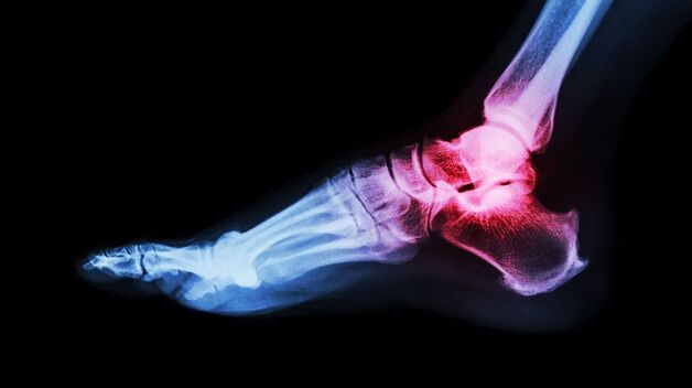
The ankle joint is often damaged due to high load. A diagnosis such as ankle arthrosis is not uncommon. They are placed regardless of the patient’s age and gender. What is ankle arthrosis and how to treat it?
Which?
There is a huge load on the ankle. Its function is to keep the body upright. Thanks to him, the man walks and runs. It is extremely difficult to lead a familiar lifestyle by violating the ankle system. What interferes with ankle work?
Ankle arthrosis, what is it? It is a chronic joint disease characterized by a degenerative course. Irreversible processes begin in the cartilage of the joint, leading to frightening complications.
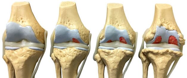
Ankle arthrosis develops gradually. Healthy joint surfaces are flexible and smooth. They provide cushioning under heavy load and smooth glide while driving. With pathology, tissue trophism and metabolism are disrupted. The surface of the joint becomes inflexible and rough. During movement, the cartilages come into contact with each other, leading to inflammation. When lifting weights, the main load falls on the bone, which threatens degenerative disorders.
Lack of treatment leads to more serious disorders. In stages 3-4, cartilage and tissue damage are observed. The synovium is inflamed. The joint becomes unstable. The support function is corrupted. All of these violations, taken together, lead to movement becoming impossible.
Arthrosis (osteoarthritis) is one of the most common joint diseases that affects quite a few people.
Causes and risk factors
We have ankle arthrosis, we arranged. Now let's see what the cause is. Ankle arthrosis is considered an pathology of old age. This is due to age-related changes in the body. The cartilage becomes thinner and the bones become unstable and brittle. In the last decade, however, the diagnosis of ankle arthrosis has become much younger. Such statistics are disappointing as many patients ignore the first signs of the disease. Late diagnosis always puts you at risk for serious complications.
Provocative factors include:
- dislocations;
- bruises;
- inflammatory diseases;
- injury;
- overweight;
- impaired metabolism;
- intolerable physical activity;
- wearing uncomfortable shoes;
- autoimmune and endocrine diseases;
- osteochondrosis.
Clinical symptoms
Ankle arthrosis is recognized by the following features:
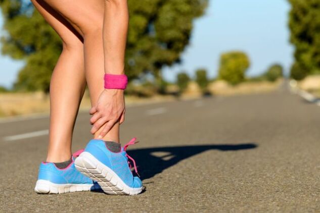
- Pain. It is mild at first and occurs after walking or physical exertion. Sometimes when a person is in an awkward position. As the pathological process progresses, the pain syndrome intensifies and you are already anxious at rest.
- Swelling and inflammation. These signs appear in the background of injuries and dislocations. Body temperature rises in the affected area.
- Click. When the ankle is affected, the click is "dry" and causes a painful attack.
- Dislocation or subluxation. Due to the thinning and degradation of the cartilage tissue, the joint becomes unstable. Bones can shift and fall out of the joint capsule. These changes cause acute pain.
- Joint stiffness. When cartilage tissue is replaced, the bone joint ceases to function normally, which negatively affects its mobility.
- Joint deformity. The symptom appears in stages 3-4 of arthrosis. Osteophytes also lead to ankle curvature.
If any of the symptoms appear, it is recommended that you see a doctor immediately. Treatment started on time is a step towards recovery.
Arthrosis of the joints of the legs and ankles is characterized by slow progression, with gradual development of clinical manifestations over several years.
Classification and sections
The disease develops in different ways. In some patients it takes several years from the first signs to the final stage, in others a rapid development of the disease is observed. The rate depends on the patient's age and state of health, the time of starting treatment. The symptoms of ankle arthrosis become clearer as the disease progresses.
Arthrosis has four stages:
- The first stage is often unnoticed. Sometimes morning stiffness and ankle pain occur after a great deal of effort. When the foot moves, a characteristic crackle is heard. X-rays do not yet show any abnormalities, but the destructive process of cartilage has already begun.
- The morning stiffness drags on. It takes 20-30 minutes for the foot to develop. Sometimes lameness occurs. Grade 2 arthrosis of the ankle joint is recognized on the radiograph by the growth of bone tissue and the displacement of the bones.
- The symptoms are expressed in 3 stages. The pain no longer worries you after a heavy load, but also at rest. The patient has difficulty living without painkillers. Lameness grows. Crutches may be required. The affected joint was swollen and deformed. The ankle muscles wither. X-ray shows narrowing of the joint space, formation of osteophytes, subluxation.
- Stage 4 is the most difficult. It develops as a result of lack of treatment. Cartilage is destroyed, the surfaces of the joints fuse. It is no longer possible to walk.
With the development of ankle osteoarthritis, there is a gradual change in the cartilage and bone tissue of the joint surfaces.
Diagnostics
The diagnosis of ankle arthrosis is based on clinical symptoms and information obtained from studies. Laboratory tests are considered ineffective because there are no specific tests that can detect pathology. During the remission period, all indicators are within normal limits, and as the disease worsens, clinical blood tests show high levels of C-reactive protein and ESR. These indicators indicate that the pathological process has already begun.
Instrumental methods are used to confirm the diagnosis:
- X-ray;
- Magnetic resonance imaging;
- Ultrasound;
- bone scintigraphy;
- diagnostic joint defect.
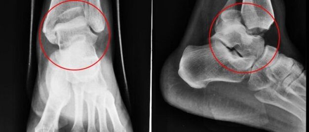
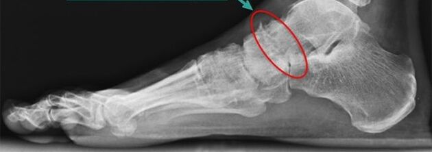
Simple X-ray
Simple X-rays are the most reliable and effective way to diagnose diseases of the musculoskeletal system. The principle of manipulation is the different absorption of X-rays by muscle tissue. Soft tissues allow X-rays to pass through, but hard tissues are absorbed. X-rays allow the diagnosis of both the disease and its consequences.
Conventional X-rays are a method of examination in which a small amount of X-rays is transmitted to a person's body or part of the body.
The snapshot allows you to view:
- The state of bone surfaces in articulation.
- The shape, size and arrangement of the structures in the joint relative to each other.
- Fabric condition.
- The size of the joint space.
These indicators help the doctor determine the type and extent of joint damage. If the data are insufficient, doctors will prescribe other tests.
In case of ankle arthrosis, X-rays are taken in three projections:
- side;
- back;
- backwards, legs moved inward.
The disease is characterized by the following changes:
- reduction of joint space;
- presence of osteophytes;
- bone cartilage replacement (subchondral sclerosis);
- smaller cavities in the periarticular part.
Nuclear magnetic resonance
Nuclear magnetic resonance (NMR) as a diagnostic method allows the study of parts of the body where there is water. The picture shows bones in dark because they contain less water, but the muscle tissue, plates, and nerves are lighter. MRI allows the detection of the slightest change in the structure of bone tissue and joints. The test is also prescribed to patients before joint prostheses. The disadvantage of YMG - high price.
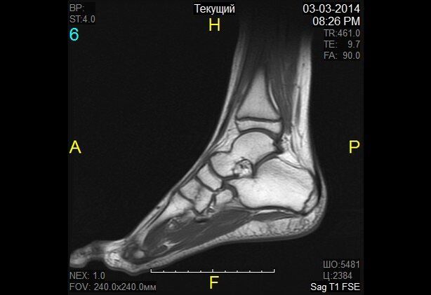
In the case of nuclear magnetic resonance, the change in the properties of hydrogen molecules under the influence of a strong magnetic field is recorded.
Magnetic resonance imaging
Magnetic resonance imaging (MRI) is an alternative diagnostic method that allows you to carefully examine the structure of the ligament structure of the joint, muscle, and cartilage tissue. An MRI is used by the doctor to assess the condition of the joints in the lower legs. Based on the survey data, the pathology is detected in the early stages of development.
The diagnostic principle is based on exposure to radio waves and strong magnetic radiation. The magnetic field used is not hazardous and does not pose a health hazard.
MRI is contraindicated in mental disorders during pregnancy and in the presence of metal objects in the human body.
Classic (closed-type) MRI machines are used to diagnose ankle arthrosis because they have better image quality. The MRI machine is a large, cylindrical tube surrounded by a magnet. The patient is lying on a special table. The ankle is secured with a special roll. The procedure takes 30-40 minutes. The test is completely painless. Patients may feel warmth in the lower leg area.
Ultrasound
Ultrasound has been widely used in medicine since the 1990s. This technique is well established in making an accurate diagnosis. Ultrasound is also performed for ankle arthrosis.
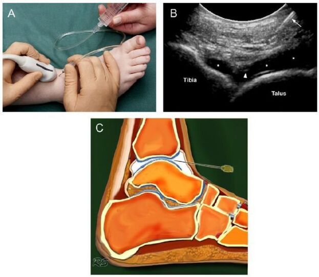
Today, ultrasound examination has no particular significance in the diagnosis of osteoarthritis because it does not allow for a sufficiently good examination of damaged joints.
The device with which the test is performed generates ultrasonic waves. The waves are reflected from the tissues and recorded on the monitor. Based on the image obtained, the doctor determines the type of pathology. In order to keep the image on the monitor clear, a special gel is used. It eliminates air gaps and provides better slip for the sensor.
Ultrasound does not harm the patient, so the procedure can be repeated many times. The benefits of ultrasound include low cost and high accuracy.
The following indicators are clear signs of arthrosis:
- cartilage thinning;
- the presence of bone growth;
- accumulation of effusion in the joint cavity (synovitis);
- loss of cartilage space.
Bone scintigraphy
Scintigraphy is a high-precision test that can use isotopes to detect abnormalities in bone. Doctors classify pathogenic foci as "cold" and "warm. "In the first case, we are talking about zones in which there are no isotopes. These areas are poorly blooded and are not visible during scanning. "Cold" areas are places that are affected by malignant tumors. In "hot" areas, isotopes accumulate quickly and are very bright when scanned. Such areas indicate the presence of inflammatory processes.
The role of scintigraphy in arthrosis is significant. The study helps to distinguish arthrosis from many other diseases when the clinical symptoms are extremely similar.
During bone scintigraphy, a special preparation containing labeled atoms is injected into the body.
Based on the results of the scintigraphy, the physician makes a clinical prognosis and determines the treatment regimen. The only disadvantage of the study is the high cost. Scintigraphy is done with special equipment and unfortunately not all health care institutions can afford to buy it.
Although radioactive scanning is a safe procedure, it still has a number of contraindications:
- pregnancy;
- lactation period;
- taking barium-containing medicines.
When radioactive material is injected, some patients experience an allergic reaction in the form of itching and rash. These side effects are not dangerous and will go away on their own in a short time.
Joint defect
A joint defect is a diagnostic procedure in which a needle is inserted into the joint cavity to collect joint fluid. This fluid is forwarded for further research. Based on the data obtained, the doctor draws conclusions about the nature of the disease and the stage of its development.
At first glance, stabbing is a simple procedure, but it is not. Removing fluid from the joint capsule requires exceptional accuracy from the physician’s movement. The synovium is very thin and traumatized by an unpleasant movement. As a result, an inflammatory process develops. Potential risks include infection. It is not difficult to introduce the infection into the joint capsule with poorly sterilized instruments.
The manipulation technique is different for each joint. When collecting joint secretion from the ankle, the puncture should be performed at the front, between the outer ankle and the extensor digitorum longus tendon.
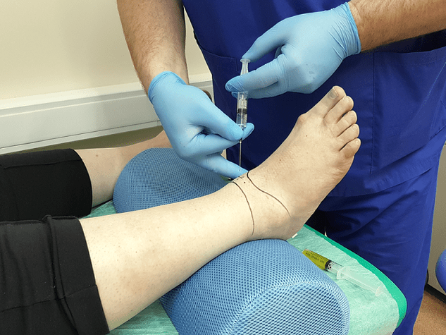
Diagnostic sampling of intraarticular fluid allows for laboratory analysis and excludes inflammatory arthritis.
Principles of treatment
Once you have confirmed the diagnosis of ankle arthrosis, the symptoms will not appear for long. Treatment begins immediately. Further prognosis depends on a well-chosen treatment regimen and timeliness of initiation.
Arthrosis is an insidious disease. It cannot be completely cured. The goal of therapy is to stop degenerative processes and prolong the period of remission. To this end, doctors prescribe medications, physiotherapy, massage, physiotherapy, and folk remedies. If all conditions are met, positive dynamics can be expected, otherwise the disease will progress.
Medication for arthrosis
Depending on the therapeutic effect, the drugs are divided into several groups:
- Anti-inflammatory or analgesic. The purpose of this class of drugs is to eliminate the focus of inflammation and relieve pain. The earlier anti-inflammatory therapy is started, the greater the chance of saving the joint. Drugs in this group can be prepared in the form of tablets and ointments.
- Glucocorticoids. These medications are prescribed when the above funds are ineffective. It is prepared as a solution for injection. The medicine is injected directly into the joint.
- Chondroprotectors. It is designed to slow the destruction of cartilage.
The treatment regimen and dosage are selected by the physician based on the severity of the symptoms, the age of the patient, the presence of concomitant diseases, and other factors. Self-medication is dangerous and often exacerbates the situation, as many medications have a number of side effects and have their own contraindications.

Characteristics of radical treatment
If conservative therapy fails, doctors are forced to take a radical method of treatment (surgery). The action also appears if:
- secondary (post-traumatic) and primary arthrosis 3-4 degrees;
- arthrosis with complications;
- constant and severe pain in the ankle, radiating towards the knee;
- severe lameness;
- paresis and paralysis of the leg muscles;
- violation of the flexor-stretching function of the joint;
- violation of the support capacity of the foot.
Surgery is contraindicated if:
- the patient is under 12 years of age;
- fistulas are found in the joint;
- the patient has diabetes, heart failure;
- Infectious diseases were found in the area of the proposed intervention.
Traditional treatment
Doctors believe that the treatment of arthrosis should be performed only under the supervision of a specialist, but do not deny the positive effect of folk remedies. Alternative medicine acts as an effective prophylaxis that helps eliminate symptoms and maintain remission.
Folk remedies are a rather symptomatic treatment for foot arthrosis.
Treatment at home should be coordinated with your doctor to avoid side effects and complications.
Traditional healers recommend treating ankle arthrosis:
- Burdock. Wash the leaves on the burdock with soap and running water. Apply the leaves with the soft side to your skin. Secure the top with a bandage or foil. It is better to keep the compress all night.
- Sea salt. Chop the salt in a pan. Pour into a canvas bag and secure to your ankles. Hold the sachet until the salt cools. The heat relieves pain. Instead of salt, sand, lentils and buckwheat are also used.
- Purple. Pour the triple cologne over the purple flowers. Leave the tincture in a dark and cool place for 10-14 days. Rub the affected area in the morning and evening.
- Eggshell. Grind the shells in a coffee grinder. Take the resulting powder on half a teaspoon. before eating.
Remember that treatment with folk remedies may not be the only measure. Complex treatment includes medication, training therapy, massage, physiotherapy, medical treatment. In advanced cases, doctors resort to radical measures - surgery.
Surgery
In arthrosis of the foot, the following types of operations are used in medicine:
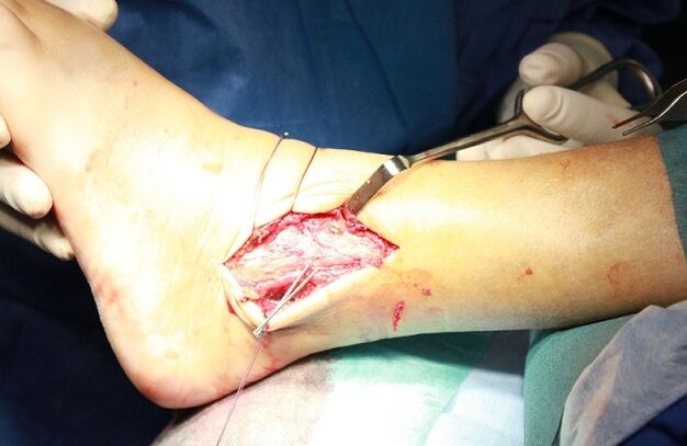
- arthrodesis of the joint;
- arthroscopy of the joint;
- endoprosthetics.
Arthrosis is an operation to immobilize the joint. This is done to restore the limb of lost support ability. The only downside to surgery is that the bones (tibia and talus) grow together, leading to immobility. Arthrodesis is rarely used in medical practice.
Arthroscopy is a minimally invasive procedure. During the surgery, the doctor makes small incisions in the joint area and inserts an arthroscope (a special tube with a camera at the end) through them. With its help, the surgeon thoroughly examines and assesses the condition of the intraarticular structures. If necessary, pieces or blood clots from the damaged joint are removed from the joint fluid. This manipulation is less traumatic. The only downside to arthroscopy is that the risk of recurrence is too high.
Endoprosthetics is the last resort. It is performed in advanced arthrosis. Endoprosthetics allow partial or complete replacement of the affected joint. As prosthetic products, prostheses with innovative, modernized mechanics are used. The artificial joint lasts for 10-20 years.
Performance characteristics
In order to achieve a favorable result, the drug treatment is supplemented with diet therapy. Nutritionists have developed a special diet to avoid the disease getting worse while providing the body with all the vitamins and nutrients it needs. The diet of overweight patients plays a special role. Because obesity is one of the causes of arthrosis, weight correction is an integral part of treatment.

The patient needs to rethink many of the habits in daily life that contribute to and provoke the progression of foot arthrosis.
Nutritionists recommend that you observe the following nutritional conditions:
- Eat often and in small portions.
- Drink at least 2 liters of fluid a day.
- Add the candy and salt.
- The last meal is no later than 18: 00.
- Steaming, cooking or baking is allowed.
The main task of the arthrosis diet is a balanced and fortified diet. Fasting is out of the question. A hard diet and body cleansing do more harm than good. Calcium is cleared from the body, which is needed to restore cartilage. A nutritionist will help you compile your daily diet.
In case of arthrosis, the consumption of cereals, pasta, dairy products, cheese, legumes, vegetables, fruits, rye bread, dried fruits, nuts, fish, poultry is allowed. Foods containing heavy and greasy garnishes, colors and flavors, as well as pickles, marinades, smoked meats, fatty broths, baked goods, spices, sauces, chocolate, ice cream, coffee and alcohol are prohibited.
Prevention of arthrosis
To prevent the development of ankle arthrosis, doctors recommend taking preventive measures:
- wear comfortable shoes without heels;
- follow a diet and drink enough fluids;
- take vitamin and mineral complexes seasonally;
- swimming;
- walk more in the fresh air;
- eliminates excessive stress on the legs;
- avoid hypothermia;
- doctor in due time.
Lifestyle correction is recommended for pre-existing arthrosis:
- Rejection of bad habits. They have been shown to provoke blood stagnation in tissues and accelerate cartilage destruction.
- Perform a series of exercises to warm your ankles.
Forecast
Arthrosis is a progressive disease. Without treatment, it leads to irreversible consequences and complete immobility of the joint. Early diagnosis of pathology allows it to be performed without radical measures. Medications have the ability to stop the pathological process and alleviate the patient’s condition. The fight against the disease takes place at an early stage without complications.